Childhood Epilepsy: Causes And Detection
Thinking about childhood epilepsy as an alteration in the quality of life is complex. The entire family of the patient with the disease is involved in their care, taking medications and modifying habits that could be harmful.
Epilepsy is a neuronal transmission disorder, in which many nerve cells perform electrical discharges at the same time. According to the location of the electrical activity, it will be the types and classification of the same. Next, we will review its causes and detection methods.
Prevalence and classification of childhood epilepsy
The data collected by the Child Neurology Foundation establish that, of all children with diagnosed epilepsy, about 40% have the disease without seizures. This is important for diagnostic differentiation, as they are not the same.
A little one can have seizures without being epileptic. If the symptom appears, the necessary complementary methods will be carried out to establish the cause, but perhaps the disorder is due to another neurological or vascular pathology.
It is also known that about 20% of epileptic children also have an intellectual disability. This affects the schooling of infants with the disorder, who must not only take care of epileptic seizures, but also see their training and literacy delayed.
But not all childhood epilepsies are the same. According to the international classification, there are some that start in the front part of the brain and others on the sides.
In addition, they can present with or without seizures, with added symptoms, with loss of consciousness or without fainting. Even within those convulsive, the attacks can be generalized or focused on a limb, for example.
Causes and risk factors
Rather than establishing an origin of childhood epilepsies, it is best to understand that there are risk factors and causal situations. The intrinsic mechanism of neuronal firing is not yet fully understood, but there are records of contexts that favor the disorder.
Within the age of infancy, the greatest risk is in the early ages. There, up to 4 years, febrile seizures can be confused with epileptic episodes. The insight of neurologists and pediatricians is critical at this point.
Genetics and family history are also important. When parents or grandparents are epileptic, a child is more likely to develop the pathology. Some genes have been classified as triggers, but they must always be stimulated by external factors that determine their expression. This means that hereditary carriage will not always result in symptoms.
Brain and meningeal infections are another cause in childhood. Many times not at the same time as the acute picture, but as a consequence and sequel of encephalitis, sometimes months and even years later.
Similarly, head injuries can lead to epileptic sequelae. This is of care in sports, especially contact exercises, such as soccer, for example. A child hit on the head can recover quickly, with full consciousness, and later be diagnosed with epilepsy.
Finally, vascular diseases form another causal group. We are referring to alterations in cerebral circulation, such as arterio-venous malformations or cerebrovascular accidents.
These disorders are not as common in children as in adults, but should be suspected if there are indicative symptoms, such as seizures or impaired mobility of the limbs.
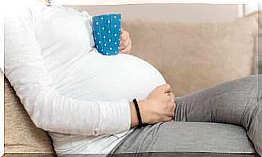
A special section is that of prenatal conditions. During pregnancy, toxins can pass from mother to fetus, affecting neuronal development. Tobacco and alcohol are two drugs that, by crossing the placenta, cause syndromes in the baby, which eventually alters electrical transmission.
How is childhood epilepsy detected?
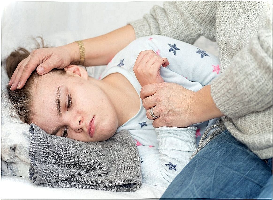
The suspicion of childhood epilepsy begins with the symptoms. In general, the first indication is a seizure that is not associated with fever in a child. From there, a series of complementary methods begin, guided by the pediatrician and the neurologist. Among these, the most important are those discussed below.
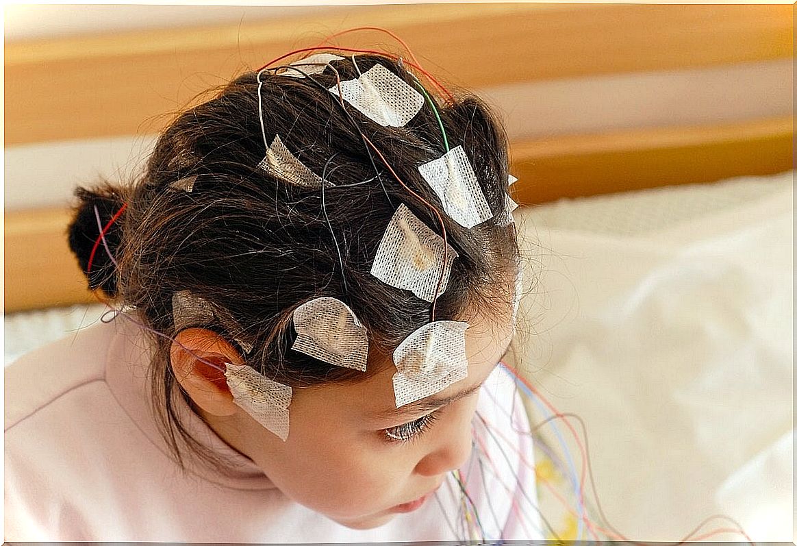
Electroencephalogram
An electroencephalogram is a test that measures the electrical activity of the brain. Electrodes are placed that, from outside the body, record neuronal transmission on a line. This record is read by specialists to know if there are anomalous discharges. It does not need to be done specifically during a seizure.
Brain scan
The computerized axial tomography (CT) of the brain is capable of detecting abnormalities in the structures of the skull that could cause seizures. A tumor, increased pressure in the cerebrospinal fluid, or an arterio-venous malforation.
Magnetic resonance
In the same way as computed tomography CT, magnetic resonance imaging is an adjunct to observe other softer structures.
Positron emission tomography (PET)
Positron emission tomography (PET) has recently gained ground as a complementary method for various pathologies, including childhood epilepsy.
By injecting a metabolic substance into the patient, which is taken up by the cells to be studied, it is possible to see this activity in the form of colors on a CT or MRI scan. This could detect tumors or neuronal areas with high activity, above what is expected, marking epileptic foci.
When in doubt, it is best to consult your pediatrician
Childhood epilepsy is not a disease that can be left unaddressed. The sooner means of treatment and prevention are established, the better the quality of life for the child and his family. Suspicion is not easy in many cases, but if a seizure does occur, professional evaluation should prevail.
The treatments are varied and have changed over the years. Today there are more specific and more sensitive diagnostic tools, as well as follow-up and rehabilitation protocols that are more accessible.